
• Histological light microscopy of bone diagenesis is a microscopic method that can be used to identify diverse funerary processes or taphonomic trajectories in archaeological human remains.
• If a body is buried whole shortly after death, the bacteria from within the body will destroy bone cells as part of the putrefaction process. If a body is treated to facilitate rapid decomposition, this process is interrupted and the osteons will be preserved.
• From this, early post-mortem treatments, as well as the type of environment decomposition occurred, can be inferred.
What is histological light microscopy of bone diagenesis?
Now more than ever, archaeology is truly a multi-disciplinary subject. Research that incorporates other fields, in this case biology, has led to the development of methods that can be applied to enhance our understanding of life and death in prehistoric societies.
The method discussed here is a histological method that shines light through thin cross-sections of human bone to measure the degree of bone diagenesis. Histology means the study of microscopic surface of tissues. Human bone often preserves in archaeological contexts, and so histological methods can be applied to study them. Bone diagenesis is defined as any taphonomic process that degrades or alters bone, leading to its destruction or fossilisation [1, 2, 3].
This post will (very) briefly explain how this analysis works and why it is important to understanding mortuary practices in archaeological populations.
What questions does it answer?
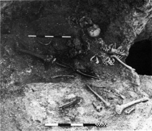
It is not always easy to determine funerary rites in prehistory. For example, during the Iron Age (c. 800 BC – AD 43) in Britain, isolated human bones, or body parts (e.g. legs, arms, torsos) are found deposited in various places throughout (and outside) a settlement. An example of isolated bones from several individuals deposited in one disused storage pit can be seen in Fig.1.
Bones can become removed from the body by a number of processes: this can be an intentional part of the burial process (e.g. excarnation, curation/circulation as a sort of relic), or an accidental disturbance (e.g. ploughing or being cut by a later grave). For the Iron Age, the most popular explanation is that disarticulated and isolated bone represents the result of excarnation practices [4, 5, 6, 7, 8].
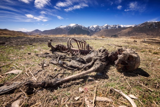
Excarnation describes a funerary process in which a body is exposed to the elements, facilitating rapid decay and skeletonisation. Excarnation is still practiced today, for example the Tibetan Sky Burial seen in Fig.2. Sometimes excarnation is followed by a secondary burial practice in which the bones are collected for redeposition in a different location.
However, it was noticed through taphonomic observations, or markers on the surface of the bone that can indicate post-mortem treatments, that the usual signs of exposure (cracking, flaking, trampling, animal gnawing) were not as common on disarticulated bone as would be expected [9, 10, 11]. A closer look inside the bone through histological light microscopy can help to solve this puzzle.
What is needed?
Interestingly, there is an absence of bacterial bioerosion from young infant skeletons, likely due to the infants dying before their osteolytic gut bacteria had developed [12, 13, 14]. Therefore, this method can only apply to adults to determine early post-mortem treatment.
- Fig.3. Anatomy of bone structure demonstrating the microstructure of compact bone. (Source)
A transverse cut of cortical bone, or compact bone, is required to assess the preservation of microstructure. Long bone midshafts, most often those from the femur, are preferred to sample for consistency. Fig.3 illustrates the anatomy of coritcal bone used for sampling. A sample needs to be large enough to create several thin sections to allow for some error—samples will often disintegrate if the cut is too fine. Results from a single long bone thin section should be representative as bacterial bioerosion in archaeological human remains does not vary significantly along long bone diaphyses [14, 15].
How does it work?
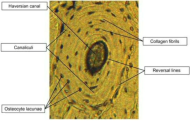
The method observes preservation of osteons, or bone cells. The anatomy of an osteon can be seen in Fig.4. Human bone is birefringent, which means that the arrangement of internal microstructure becomes more visible under polarized light [3].
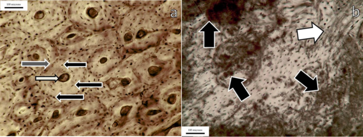
Evidence suggests that bacteria found within the gut (endogenous gut bacteria) may be involved in the destruction of the osteons [12, 16, 17, 18]. During the first few days after death, visceral bacteria, or bacteria that occurs naturally in the gut, transmigrate around the body via circulatory system. Poor preservation of bone microstructure is typical of articulated burials [18, 19], when the bacteria is able to attack the bone cells over a longer period of time. Conversely, good preservation suggests that decomposition of the soft tissue occurred more rapidly and inhibited the bacteria from accessing the bone, for example excarnation by subaerial exposure (e.g. Fig.2). The difference is illustrated in Fig.5.
The bioerosion is measured using the Oxford Histological Index (OHI) as seen in Fig.6. The OHI is an ordinal measure of bioerosion relating to the percentage of unaltered bone microstructure [1, 3, 20].
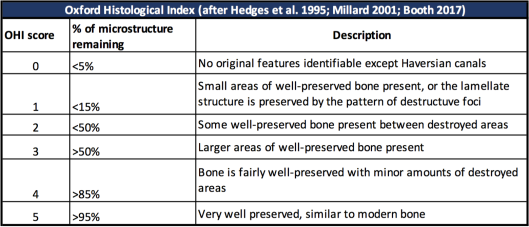
- An OHI score of 0-1 would suggest that a corpse was buried whole and left to decay slowly in the ground. This is typical of inhumation grave burials.
- An OHI score of 2-3 is a tricky middle score that can mean that the body was in an environment that facilitated quicker-than-usual decomposition, but not exposure. A possible explanation is that the body was placed in a covered pit or shelter.
- An OHI score of 4-5 indicates that soft tissue was removed from the skeleton rapidly. For example, rapid skeletonisation could be the result of subaerial exposure or human processing/defleshing.
What does it mean?
Funerals were often complex in prehistory, requiring several phases and multiple rites. As mentioned, in the Iron Age, this is suggested by the skeletal remains in various states of completeness found within various features, including grain storage pits, boundary ditches surrounding a settlement, post holes that hold the large timber frames of residential houses, as well as caves, rock shelters and specially cut graves. We can make observations about secondary treatments of these disparate bones by analysing macroscopic, or surface taphonomy. However, in order to understand the primary treatments, you have to go deeper.
Was the person buried whole, only to be disturbed and reburied at a later date? Was their body exposed to the elements and the bones picked clean by carnivorous animals? Or was it something in-between, like exposure in a protected environment such as a covered pit? A closer look inside the bone through histological light microscopy of bone diagenesis can help determine what happened to the body in the early post-mortem period.
This has challenged the preconceived notions around Iron Age burial practice. It appears that there were more rites afforded to different classes of bone than previously realised. Instead of excarnation by subaerial exposure being the majority rite, it is more likely that it was one of many options for disposing of a corpse and seeing a person through to whatever lies beyond the mortal world. As more data is collected on the diagenesis of archaeological populations, coupled with established taphonomic methods, a more holistic understanding of ancient funeral rites will be gained.
Bibliography
[1] Hedges, R., Millard, A., and Pike, A. 1995. Measurements and relationships of diagenetic alteration of bone from three archaeological sites. Journal of Archaeological Science 22, 201-209.
[2] Lee-Thorp, J., and Sealy, J. 2008. Beyond documenting diagenesis: the Fifth International Bone Diagenesis workshop. Palaeogr. Palaeoclimatol. Palaeoecol. 226, 129-133.
[3] Booth, T. 2017. The rot sets in: Low-Powered Microscopic Investigation of Taphonomic Changes to Bone Microstructure and its Application to Funerary Contexts. In T. Thompson and D. Errikson (eds.) Human Remains: Another Dimension. Chapter 2, 8-28.
[4] Ellison, A., Drewett, P.L. 1971. Pits and post-holes in the British early Iron Age: some alternative explanations. Proceedings of the Prehistoric Society 37, 183-184.
[5] Stanford, S.C. 1974. Croft Ambrey, Excavations Carried Out at the Woolhope Naturalists Field Club 1960-66. Woolhope Naturalists Field Club, Hereford.
[6] Dunning, G. 1976. Salmonsbury, Burton-on-the-Water, gGoucestershire. In: D. Harding (ed.), Later Prehistoric Earthworks in Britain and Ireland. London: Routledge, 75-118.
[7] Carr, G., Knüsel, C. 1997. The ritual framework of excarnation by exposure as the mortuary practice of the early and middle Iron Ages of central southern Britain. In: A Gwilt and C. Haselgrove (eds.), Reconstructing Iron Age Societies. Oxford: Oxbow Monograph 71, 167-173.
[8] Cunliffe, B., Farrell, P., and Dee, M. 2015. A happening at Danebury Hillfort, but when Oxford Journal of Archaeology 34, 407-414.
[9] Madgwick, R. 2008. Patterns in the modification of animal and human bones in Iron Age Wessex: revisiting the excarnation debate. In: O. Davis, N. Sharples, and K. Waddington (eds.), Changing Perspectives on the First Millennium BC. Oxford: Oxbow, 99-118.
[10] Madgwick, R., 2010. Bone modification and the conceptual relationship between humans and animals in Iron Age Wessex. In: J. Morris and M. Maltby (eds.) Integrating Social and Environmental Archaeologies: Reconsidering Deposition. Oxford: BAR International Series 2077, pp. 66-82.
[11] Booth, T. and Madgwick, R. 2016. New evidence for diverse secondary burial practices in Iron Age Britain: a histological case study. Journal of Archaeological Science 67, 14-24.
[12] White, L., and Booth, T. 2014, The origin of bacteria responsible for bioerosion to the internal bone microstructure: results from experimentally-deposited pig carcasses. Forensic Science International, 239, 92–102.
[13] Booth, T. 2016. An investigation into the relationship between bacterial bioerosion and funerary treatment in European archaeological human bone. Archaeometry 58(3), 484-499.
[14] Booth, T., Redfern, R., and Gowland, R. 2016. Immaculate conceptions: micro-CT analysis of diagenesis in Romano-British infant skeletons. Journal of Archaeological Science 74, 124-134.
[15] Del Sasso, G., Martian, L., Usai, D., Angelini, I., and Artioli, G. 2014. Bone diagenesis at the micro-scale: bone alteration patterns during multiple burial phases at Al Khiday (Khartoum, Sudan) between the Early Holocene and the II century AD. Palaeogeogr. Palaeoclimatol. Palaeoecol. 416, 30-42.
[16] Child, A.M. 1995. Towards an understanding of the microbial decomposition of archaeological bone in the burial environment. Journal of Archaeological Science 22, 165-174.
[17] Bell, L., Skinner, M., and Jones, S.1996. The speed of post mortem change to the human skeleton and its taphonomic significance. Forensic Science International 82, 129-140.
[18] Jans, M.M.E., Nielsen-Marsh, C.M., Smith, C.I., Collins, M.J., and Kars, H. 2004. Characterisation of microbial attack on archaeological bone. Journal of Archaeological Science 31, 87-95.
[19] Nielsen-Marsh, C.M., Smith, C.I., Jans, M.M.E., Nord, A., Kars, H., and Collins, M.J. 2007. Bone diagenesis in the European Holocene II: Taphonomic and environmental considerations. Journal of Archaeological Science 34 (9), 1523-1531.
[20] Millard, A. 2001. The deterioration of bone. In: D. Brothwell and A.M. Pollard (Eds.), Handbook of Archaeological Sciences. Chichester: John Wiley & Sons, 637-647.
Other:
Fig.1. Cunliffe, B. 1991. Danebury, An Iron Age Hillfort in Hampshire Vol 5: The excavations 1979-1988: The Finds. York: CBA Research Report no. 73

Contributor
Deputy Editor, Adelle Bricking
Adelle is a postgraduate research student at Cardiff University. She studies Iron Age mortuary practices in southwest Britain.
Reviewed by: Eirini Konstantinidi

Leave a comment